Hematuria
Compiled case information:
Mr. A is a 56-year-old man who has had several episodes of red urine in the past few days.
Mr. A reports several episodes of painless visible hematuria over the last several days, along with occasional mild lower abdominal discomfort. He is feeling well otherwise and has no other complaints. His medical history is significant for chronic kidney disease (CKD) stage 3, hypertension treated with hydrochlorothiazide and enalapril, and a remote appendectomy. He has no family history of kidney stones, but his father did have prostate cancer. He has smoked 1 pack per day of cigarettes for 35 years. He is a philosophy professor, and has no known toxin exposures. Initial urinalysis shows many nondysmorphic RBCs, with no WBCs, bacteria, casts, or proteinuria.
Mr. A’s physical exam is normal, with no abdominal masses or tenderness. External genitalia are normal, and digital rectal exam shows a symmetric, nontender prostate without nodules. Serum creatinine is 1.8 mg/dL, unchanged from previous values.
Urine culture is negative.
Although you are concerned about malignancy, because of his CKD you order an ultrasound rather than a CT scan. It shows a 1 mm stone in the right renal pelvis, and a 2 cm cyst in the left kidney. You order a PSA and refer him to urology for a cystoscopy.
Mr. A’s cystoscopy detects a small papillary tumor localized to the uroepithelium of his bladder. Hexaminolevulinate fluorescence cystoscopy does not detect any carcinomas in situ. CT urography does not demonstrate any masses elsewhere in the upper urinary tract or kidneys. His bladder cancer is classified as superficial and he is treated with transurethral resection followed by BCGtherapy. At a follow-up visit 1 year later he is cancer free and feeling well.
Soap note:
S: 56 y/o male complains of several episodes of painless visible hematuria over the last several days, along with occasional mild lower abdominal discomfort. He has significant past medical history of chronic kidney disease (CKD) stage 3, hypertension treated with hydrochlorothiazide and enalapril, and a remote appendectomy. His father had prostate cancer. He has smoked 1 pack per day of cigarettes for 35 years. No known toxin exposures.
O: Abdomen/Genitourinary System/Rectum: No abdominal masses or tenderness, external genitalia unremarkable, and a symmetric, nontender prostate without nodules in digital rectal exam.
Urinalysis: many nondysmorphic RBCs, with no WBCs, bacteria, casts, or proteinuria. Urine culture: negative. Serum creatinine: 1.8 mg/dL.
Ultrasound: a 1 mm stone in the right renal pelvis, and a 2 cm cyst in the left kidney. Cystoscopy: a small papillary tumor localized to the uroepithelium of his bladder. Hexaminolevulinate fluorescence cystoscopy: no carcinomas in situ detected. CT urography: no masses elsewhere in the upper urinary tract or kidneys detected.
A: Superficial bladder cancer
P: Transurethral resection followed by BCG therapy
Follow-up in 1 year
Summary:
In this case, bladder cancer is the leading hypothesis. It accounts for 90% of urothelial cancers and 85% of patients show intermittent visible painless hematuria. The related risk factors include male sex, smoking and age > 40. Studies demonstrated that white males have much higher possibility to develop bladder cancer than white females. It is also shown that 60% of bladder cancers in males and 30% in females are related with smoking. In addition, the median age at diagnosis of bladder cancer is 70. Besides, 3.7% of patients with microscopic hematuria was found to have bladder cancer. Mr. A is a 56 y/o male with several episodes of painless visible hematuria over the last several days and his initial urinalysis shows many nondysmorphic RBCs without WBCs or bacteria, so the leading hypothesis is bladder cancer. Differential diagnosis includes bladder, ureter or kidney stone, BPH, prostatitis, prostate cancer and renal cell carcinoma.
To diagnose bladder cancer, white light flexible cystoscopy with biopsies is the gold standard and is required for patients older than 40 years old with hematuria. Other useful exams include hexaminolevulinate fluorescence cystoscopy for carcinoma in situ, multiphasic CT urography with and without contrast including imaging in the excretory phase and ultrasound. Although CT urography has higher sensitivity and specificity than ultrasound, ultrasound was firstly performed due to his history of chronic kidney disease (CKD) stage 3. In addition, urinalysis and urine culture were performed to detect or exclude UTI.
Source: An Evidence-Based Guide, 3e by Scott D.C. Stern, Adam S. Cifu, Diane Altkorn
reflection
It is a great and new experience for me to write SOAP notes. I learned hematuria and bladder cancer before, but I never wrote a SOAP note. Through studying from textbook and online materials, I completed the assignment. I found that SOAP note is to put most relevant information together in a well-organized format to give a general acknowledge of the patient’s condition. I really enjoyed the process from knowing little to being confident. Through this work, I am familiar with SOAP notes and made a SOAP note that met standards of examples in the textbook. Through I am satisfied with my work, I believe that I need to practice to make SOAP notes more concise and relevant. By the way, I found that the textbook and online materials are very helpful, especially the textbook. I would use all of the sources again in my next work.
bladder cancer image
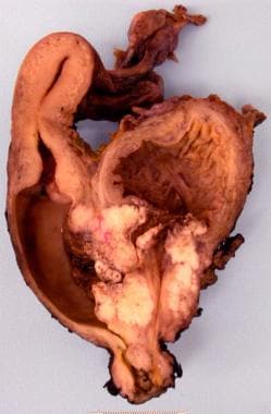
Source: https://emedicine.medscape.com/article/438262-overview

This entry is licensed under a Creative Commons CC0 1.0 Universal Public Domain Dedication license.